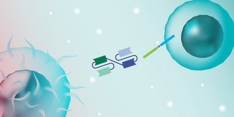
Bispecific Antibodies and Cancer Immunotherapy
Bispecific antibodies (bsAbs) are an important addition to the immuno-oncology toolbox. Designed to recognize two distinct epitopes, bsAbs have enhanced binding, specificity, and efficacy compared to current monovalent antibody therapeutics, making them exciting candidates for more targeted cancer treatments. Learn about bsAbs and how our tools can help scientists with their research on therapeutic candidates.
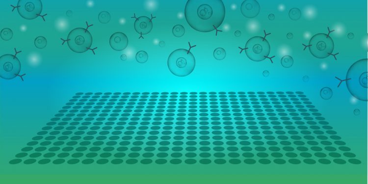
5 High-Throughput Screening Applications Using Flow Cytometry
For many scientists, high-throughput flow cytometry screens are associated with drug or compound libraries, but did you know that you can also apply this technique to study microorganisms, clinical melanoma samples, and even protein trafficking? Read on for five examples of the use of flow cytometry in diverse, high-throughput screening applications.

Advanced Cancer Therapeutics: Increasing Efficacy and Specificity and Reducing Toxicity
Research into human diseases has led to the development of novel monoclonal antibody drugs. These monoclonal antibody therapies have been successful because of their specificity and selectivity. However, ongoing development and successfully launching a new mAb drug require robust and reliable methods to screen for drug targets, demonstrate drug safety and efficacy, and scale up manufacturing to meet the stringent regulatory guidelines required for approval.

ArticlesCell BiologyCOVID-19Drug Discovery and DevelopmentFeatured StoriesGeneral InterestResearch Highlights
Immune Surveillance in SARS-CoV-2 Vaccine Development
The ability to characterize the target of cell-mediated immune response is crucial during vaccine development. In studying the characterization of SARS-CoV-2 infection pathogenesis in humans, a research team identified neutralizing antibodies that provided context for vaccine antigen design. Discover how the ZE5 Cell Analyzer was used to detect activation-induced markers, measure intracellular cytokines, and identify spike protein–specific antibodies in a rapid, high-throughput manner.

A 16-Color Flow Cytometry Panel to Monitor Checkpoint Inhibitor Expression and T-Cell Exhaustion Following Treatment with the PD1 Inhibitor Nivolumab
Presented by: Angie Green, PhD, Senior Scientist
View on demand
We discuss using a multiparameter flow cytometry panel to monitor T-cell exhaustion states and the expression of checkpoint inhibitors. We used an in vitro model; however, the panel is designed to be compatible with clinical samples to monitor immunotherapy.
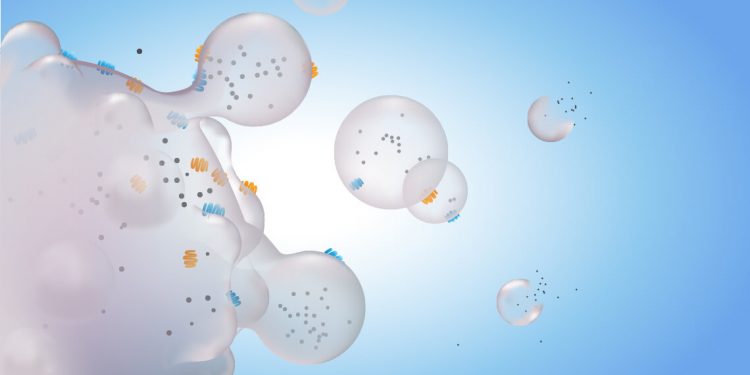
Flow Cytometry — A Gateway for Discovering More about Small Vesicles
Small vesicles were identified over 50 years ago but scientists are still trying to understand their biology and role in disease. These vesicles have become relevant for monitoring, diagnosing, and treating complex diseases such as cancer. Yet, because of their small size compared to cells, they require intricate technologies to truly understand their impact in research. Learn about the various technologies used to study small vesicles and discover the power of flow cytometry for learning more about these valuable vesicles.