
Assessing Cell Health: Apoptosis
In this second part of the series on assessing the health of your cells, we review the different types and stages of apoptosis, and the various methods of assessing apoptosis of cells.
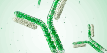
Macrophage Polarization — An Overview
Macrophages are important players in the phagocytosis process in the immune system. In this detailed review, we provide an overview on the different types of macrophages, the signaling molecules involved in the polarization of M1 and M2 macrophage subsets, and more.
Assessing Cell Health: Viability and Proliferation
In this multi-part series on assessing the health of your cells, we review different factors to be considered in your assessments such as cell viability, proliferation, apoptosis, and autophagy. The first part covers the methods for assessing viability and proliferation and their advantages, and discusses some common considerations.
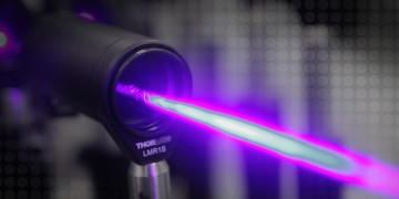
Expanding Flow Core Capabilities by Shifting Routine Sorts to Self-Sort on the S3e™ Cell Sorter
To run a core flow cytometry lab is no easy task. There is always a balance between running simple sorts and developing new assays for answering complicated questions that demand pushing the limits of existing assays and the limitations of the sorters. See how Andy Riddell, a flow core manager, pushes the limits of his existing assays and how Bio-Rad’s S3e™ cell sorter is allowing him to accomplish that.

Flow Cytometry: Basic Definitions
It’s always good to get the basics straight. Here, we’ve defined some flow cytometry terms that might have eluded you.

10 Tips and Tricks for Designing Multicolor Flow Cytometry Panels
Multiplexing an assay comes with its own complications in addition to the benefits it affords. Designing multicolor panels for flow cytometry analysis entails several considerations for a successful outcome. Here is a list of tips that can help you succeed in setting up and running a multicolor flow cytometry panel.
Protocol: Preparation of Cells for Flow Cytometry
Harvesting of tissue culture cells could be challenging. Here is an easy protocol for efficient harvesting of cells for flow cytometry analysis.