Introduction

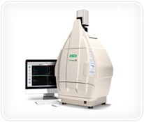
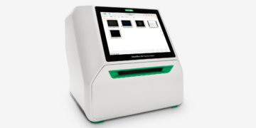
True to its long tradition of innovation in imaging, Bio-Rad has recently introduced the stain-free gel electrophoresis workflow which was rapidly followed by the launch of the Gel Doc™ EZ imager, bringing full-fledged, publication-quality imaging capabilities to a simple push-button platform. This tradition continues today with the release of the ChemiDoc™ MP imaging system, a full-feature instrument for gel or western blot imaging designed to address multiplex fluorescent western blotting, chemiluminescence detection, and general gel documentation applications.
The ChemiDoc MP system offers high-resolution CCD imaging, high-sensitivity detection technology, and modular options to accommodate a wide range of samples and support multiple detection methods. The system is controlled by Image Lab™ software to optimize performance for fast, integrated, and automated image capture and analysis of various samples. Launched globally in the fall of 2011, the system’s unique features — including easy-to-operate software, multiplex fluorescence, and utilization of the stain-free work flow — have already facilitated progress in research laboratories around the world.

Graduate Student
Molecular Physiology Integrated Graduate Program
Gomes Laboratory, University of California Davis

Shannamar Dewey, a graduate student at UC Davis, using the ChemiDoc MP imaging system. The system has cut her imaging time in half and allowed her to visualize her gel and the blot during different stages of western blotting.
Davis, California — Stain-Free Simplicity
A former student athlete, Shannamar Dewey enrolled in the molecular physiology integrated graduate program at UC Davis because she was looking to gain an in-depth understanding of the science behind exercise health and human performance. This program requires students to rotate through several laboratories to develop insight into what they are interested in and want to focus on. “Davis was the only program I saw that allows you to explore options in your first year rather than requires you to know going into your graduate program what you want to specialize in,” says Dewey. Her first rotation was into was the Aldrin Gomes laboratory, and after additional rotations, that is where she returned to complete her graduate studies. “This lab lets me do exactly what I wanted in terms of building understanding and skills in molecular physiology research at the cellular level,” says Dewey.
The primary focus of research conducted in the Gomes laboratory is on how protein degradation pathways affect cellular performance. Researchers study functional consequences of protein mutations, for example troponin mutations, and their link to sudden death in cardiomyopathy. “My work is a spin off from these studies,” explains Dewey. “We became interested in diabetic cardiomyopathy because studies have shown that there could be a role for protein degradation in the pathogenesis of that disease.” The questions she is seeking answers for include: How can protein degradation, or the regulation of protein degradation specifically through the ubiquitin proteasome system (UPS), possibly be playing a role in the pathogenesis of diabetic cardiomyopathy?
“To answer this question,” says Dewey, “we need to know if the UPS is being altered during the disease state, and if that alteration is happening pre- or post-disease.” To do this, they run assays to look at the activity levels of different proteases to see if these are being altered. Running gels is a critical basic step in their workflow as this allows them to determine how much protein is present in samples, look at protein amounts for various control versus experimental groups, and equalize protein amounts across these groups prior to running their assays. “The model I’m using is a mouse model,” says Dewey. “My samples are whole heart homogynates, and so far we’re seeing changes over time with what’s happening with the UPS.”
Imaging comes into play in Dewey’s workflow at multiple steps. “Probably the most often used step for imaging in my research is with western blotting,” she explains. “I rely on imaging to tell the true story of how much antibody is present in my control versus experimental animals.”
But prior to acquiring the ChemiDoc MP system and ability to perform stain-free imaging, Dewey found western blotting challenging, experiencing uncertainty as to whether her proteins separated well in her gels and if all her proteins transferred completely. “With the ChemiDoc MP system’s preprogrammed stain-free setting, it’s a 2 minute, 30 second activation of the stain-free gels,” says Dewey. “This allows me to be confident before I even do my transfer that my gel ran well — now I can just doublecheck my transfer before I waste my time and my antibodies.”
Elimination of the background optimization step is another time-saving feature Dewey appreciates with the ChemiDoc MP system, estimating that it has essentially cut their imaging time in half.
“My goal is not to be a graduate student forever,” says Dewey. “So in that respect, anything that makes my day faster and I’m able to fit more in — it fits into my plan.”

Assistant Professor of Biological Sciences
Korea Advanced Institute of Science and Technology (KAIST)
Korea

Graduate students in Dr Ji-Joon Song’s lab in the Korea Advanced Institute of Science and Technology find the ChemiDoc MP imaging system very useful because of its ability to image and also quantitate chemiluminescent western blots.
Daejon, Korea — Eliminating Training Pain Points
In the corner of Dr Ji-Joon Song’s office shoved between file cabinets and a wall is a stack of sleeping bags. When asked what these are for, the Assistant Professor of Biological Sciences at Korea Advanced Institute of Science and Technology (KAIST), laughs and explains they are for the students who often work long hours in his laboratory, so they can sleep onsite rather than waste time traveling to and from home. As one of the top research centers in the world, KAIST draws the top students from both Korea and abroad. “We have a really huge pool of excellent students that we can rely on to help support our research projects,” says Song.
Back when he himself was a student, Song had no intentions of becoming a scientist. In fact, he originally studied to become an architect. “I was so fascinated by how 3-D structures are constructed,” says Song, “so I knew that I was going to be an architect, not a biologist.” But it was in high school biology when he was exposed to the machinery of cells inside the body that this fascination transferred to the realm of biological sciences. These days, books on architecture and building are scattered amongst the more expected peer-review scientific journals and protocol manuals in the bookshelves that line his office walls. But Song did not leave this interest in structures entirely behind. Instead, he has merged this early life passion with his scientific career, applying the principals of structural biology to the mechanisms of biochemistry and molecular biology.
Song has spent the past two and a half years setting up his laboratory at KAIST to apply structural biology tools to investigate biochemistry and cell biology-based questions. His primary field of focus is epigenetics, specifically chromatin biology. He explains that the biggest question he seeks to resolve is how internal molecular expression patterns and machinery engage to individualize human beings who start with identical genetic material. He believes that the answers to this fundamental question have the potential to greatly improve disease prevention and treatment, specifically diseases such as cancer and Huntington’s disease. “Although cancer and Huntington’s seem to be completely different diseases,” says Song, “we are finding that they share a common epigenetic modification. My research focuses on the mechanics of this epigenetic regulation, so provides a platform for therapeutics for both diseases.”
Song explains that the most challenging part of his work is the science itself. “Nature hides so many things,” he says. “Our job as scientists is unveiling what is there. It’s not easy.” As Song continues to expand his research beyond identifying chromatin structures and into mechanisms and functions he faces an additional challenge, which is acquiring molecular and cellular biology-based tools and then training his staff and students on the use of these tools.
Common laboratory techniques in his lab would include PCR, electrophoresis, immunoblots, crystallizations, and protein purifications. Song estimates that they run 20-–30 gels per day. When determining which instrumentation to purchase to image these gels, Song considered two different options: scanning and CCD-based imagers. “We tried Bio-Rad’s ChemiDoc MP system for one week,” says Song, “and we discovered it is very user-friendly, the software very straightforward.” He states that other machines he’s used in the past require training; this one he could figure out on his own. Plus a major factor in his decision to go with the ChemiDoc MP system is the ability to use the instrument to perform quantitation; scanning technology is not practical to use for this purpose. And in terms of sensitivity? “I think sensitivity is comparable to x-ray film,” says Song, “particularly for the range that I want to detect.”
But perhaps the greatest advantages of this tool for his lab is the intuitiveness of the software. Because his lab does rely heavily on student assistance with laboratory protocols, the fact that the ChemiDoc MP system can be used to conduct a variety of imaging tasks, from the simple to the complex, with little or no training required had a large impact on their productivity.
“Now with this ChemiDoc in my lab I can do a lot of different things,” says Song. “Not just investigate the structure of molecular machinery, but I can perform experiments that can be monitored in real time to show quantitatively, how this machinery functions in terms of biochemical properties.” And he — as well as his staff and students — can accomplish these investigations without needing to invest significant time and resources in imaging-related training.

Department of Animal Physiology
University of Erlangen
Erlangen, Germany

Researchers in Dr Andreas Gießl’s laboratory at the University of Erlangen in Erlangen, Germany value the ability of the ChemiDoc MP imaging system to quantitate multiplex fluorescent western blots, especially because they have an extremely limited sample size.
Erhlangen, Germany — Color Vision Evolved with Multiplex Fluorescence
Dr Andreas Gießl’s laboratory in the Department of Animal Physiology at the University of Erlangen in Erlangen, Germany is known for its expertise with microscopes and imaging instruments in general. In particular, Gießl has fostered an expertise in electron microscopy, an expertise that only a few laboratories around the world can claim. This attention to rendering the invisible visible is interesting given the focus of the laboratory’s research: sensory systems, in particular, the eye. “We think seeing is a normal thing we can do,” says Gießl. “We can see in dark and bright light, and the eye adapts automatically to every condition.” But, he goes on to explain, most of us take these abilities for granted – except those who are not able to see. And many diseases related to the eye can be fatal.
Vision begins in photoreceptor cells, what Gießl describes as “one of the best known cells in science,” specifically for its signal transduction cascade that is known, but not yet well understood. Signal transduction takes place in the outer segment of photoreceptor cells, but the production of proteins necessary for signal transduction takes place in the inner segment where the nucleus is located. These proteins move from the inner to the outer segment of photoreceptor cells — the only intracellular connection between these parts or compartments of the cell is the connecting cilium. Ciliary structures are the primary focus of Gießl’s work. “You have many cilia distributed throughout the whole body,” says Gießl, “All cells can build cilia, and there are many diseases known, mostly development-related diseases, which are associated with these ciliary structures. In photoreceptor cells, mutations in ciliary proteins lead, in most cases, to photoreceptor degeneration.”
One of the proteins Gießl is working with is a scaffold protein that is thought to be involved in the organization of translocation processes in photoreceptor cells, and also perhaps in several diseases, like ciliopathies, polydactylism , and dwarfism. “We are merely at the beginning of understanding this protein,” says Gießl. “We can now say where it is localized in the body — in the centrosomes and ciliary structures in general and in the connecting cilia of the photoreceptors in particular — and this is brand new information.”
To answer their primary research questions, the laboratory uses a variety of tools and techniques. They perform many laboratory techniques related to these studies like immunocytochemical studies at the electron- and light microscopical level, qPCR, western blotting, ELISAs and many others. But what has proven most challenging has been devising a method to quantify protein expression levels — necessary in their work to define for example the role of their proteins of interest in different stages of development of the retina. “You can do it,” says Gießl, “but it has always been difficult with the tools we’ve had available in our lab.”
For example, a routine part of post-graduate student Johanna Mühlhans’ work centers on extracting proteins from retinal tissue of different postnatal stages of mice to observe protein patterns at different stages of development. Starting at day 0 to day 31 after birth she has observed distinct expression patterns of different proteins that alter during retinal development. “It’s a really important point for development,” says Mühlhans, “especially in mouse models that carry a knock out or knockdown of a protein. You can in many cases, for example, measure and show that different proteins compensate for this knocked out protein.” She goes on to describe limited resources as a serious constraint — when using mice of different stages of age, there are obviously limited mice of one age available for one experiment, so you have to work with a very small sample pool to represent the different ages.
To help address this limitation in their workflow, Gießl’s laboratory acquired the ChemiDoc MP imaging system. The system features multiplex fluorescence imaging, so researchers can image several proteins in the same western blot or even in one lane of the blot. “This is very helpful for us in different experimental approaches,” explains Gießl, “because when we for example downregulate a protein we can look in the same lane at the interaction partners to see if they’re also downregulated or if they’re upregulated — we can use several antibodies in one lane because of the possibility to use different colors.”
Mühlhans is also enthusiastic about this capability. “When you don’t have much material, you simply cannot do so many blots and run so many gels just to test all the proteins one after another,” she says. “The excellent thing about this ChemiDoc MP system is I can take my one blot, even from this one animal if this is needed, and I can show all the proteins I’m interested in in one lane. I don’t even need the whole blot, I just need one lane to show in different colors the expression of different proteins – and I can quantify against a host protein, for example β-actin.”
Mühlhans likens the development of multiplex fluorescence imaging capability to the evolution of vision. “In the field of vision,” she says, “we have to think about a lot of different stages of vision, starting from the very basic vision of light in general to the bright color vision of a monkey or a human being. And so I guess it’s a little bit alike with these instruments. This new ChemiDoc MP system has evolved a kind of color vision — the possibility to image these different colors is, as in nature, a great step in the evolution of our research.”


